![]()
|
|
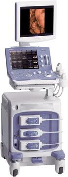
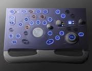
Scanner Equipment- Hitachi Aloka Alpha 6 Color & 3D ScannerThe Hitachi-Aloka Alpha6 Imaging System
Alpha 6 Imaging Modes
Alpha 6 Features Probe Frequency: Multi-Frequency- 5.0 + 7.5 + 13.0 Mhz. Here you get it all penetration and high resolution. Free Hand 3D- Optional- Give you a 3D image of tissue and masses used to evaluate and explain diagnosis to patients. File Management- Stores all your images - Up to 40,000 by name, date, and file number. USB Memory Key- Export images form the PICO to your computer through the USB Port for archiving / telemedicine. Trapazoidal Imaging- Allows a wide field view using the high resolution 12 Mhz. Linear array probe for musculoskeletal imaging. Color Doppler- Enables duplex color vascular exams, instantly distinguish arterial vs. venous, blockages, and tumor vascularity. Soft Tissue Harmonics- Matches the frequencies for dynamic soft tissue imaging to provide optimized image quality. Variable Frequency Probe- 5- 13 Mhz. Broad band to allow the operator to choose the optimal frequency for each image. 100% Digital System- The most advanced technology for image optimization and connectivity. Archiving / Telemedicine- Internal file management. export via email, MO Drive, CD, DVD, USB Stick, Video Printer, Lan SVHS Video, audio, MIC, and foot switch ports on back of the system. DICOM 3.0 (Optional)- Allow you to interface with any hospital network for individualized connectivity.
Pre-set - APPLICATIONS Musculoskeletal (foot & ankle, shoulders, wrists, hips, knees) Small Parts - General Ultrasound Vascular - Arterial and or Venous Measurements - 2D Distance - 2D Ellipse - 2D Trace - 2D Hip Joint 3D Volume (3 distance) 3D Volume Click HERE for Information/Contact Form
|
Questions? CLICK TO ZOOM IN Hitachi Aloka Alpha 6
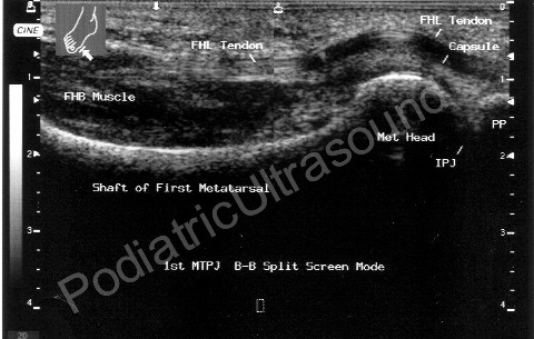
Scanner Images
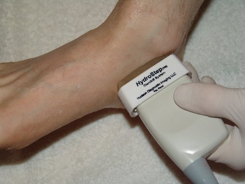
Pico Podiatric IMAGES HydroStep® Standoff
|
| Copyright © 2004 -
BioVisual Technologies LLC-
|


